3D printed mini tumors aid cancer treatments

Scientists have developed a method for ‘bioprinting’ minitumors, designed to mimic the function and architecture of real tumours. Its purpose is to help predict how patients will respond to cancer treatment.
For those in a hurry:
The process – developed by researchers at the Jonsson Comprehensive Cancer Center at UCLA (University of California) – allows for the use of an advanced imaging method to study and analyze individual organoids in detail. That’s how they help researchers identify personalized treatments for people with rare or difficult-to-treat cancers.
Tumor organoids have become key tools for studying tumor biology and highlighting patient drug sensitivities. However, we still need better ways to predict whether resistance might emerge in a small population of cells, which we can’t detect using conventional screening approaches. This is really important, particularly as organoid-based drug predictions are starting to be exploited clinically.
Alice Soragni, assistant professor in the department of orthopedic surgery at the UCLA School of Medicine
minitumors

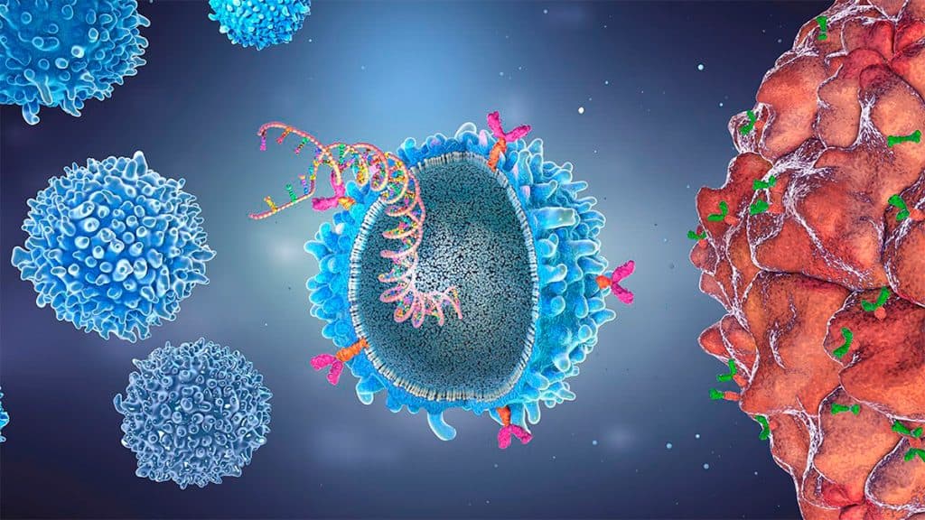
Called organoids, they can be grown in the lab using cell lines or patients’ own cells to better understand human biology and disease. By recreating tumors from patients, researchers can test different drugs to see if the tumor will respond well or poorly to treatment. This can make it easier for doctors to choose the best therapy for their patients.
These minitumors have helped improve drug modeling and are becoming valuable tools for testing the efficacy and safety of potential drugs. But it is still a challenge for current models to capture the underlying tumor heterogeneity that often leads to the clinically observed resistance to therapy.
One major limitation of this approach is that current methods fail to capture changes or differences in organoid samples that might be responsible for the therapy resistance observed in the clinical setting.
How they 3D printed ‘tumors’
To overcome these challenges, the team of researchers created a method that uses a bioprinting technique to print cells on a thin layer of extracellular support proteins to give rise to 3D minitumors without changing tissue histology and gene expressions. .
The team combined bioprinted cells with high-throughput live cell interferometry (HSLCI). This imaging system is a non-destructive approach used to observe and measure the weight of live cells in real time. The methods are combined with machine learning algorithms to analyze and measure individual organoids.
Using this method, we are able to accurately measure the masses of thousands of organoids simultaneously. This information helps identify which minitumors are sensitive or resistant to specific therapies, which can be used to quickly select the most effective treatment options for patients.
physician Michael Teitell, director of the UCLA Jonsson Comprehensive Cancer Center and co-senior author of the study
With the new combination of methods, the researchers confirmed they could measure the growth patterns of ‘bioprinted’ cancer cells over time to see how the cells respond to different drugs or treatments.
Search Results

The researchers were able to identify an effect of some drugs on the cells six hours after the therapies were added. The team also identified small clusters of cells that were unresponsive to the drugs, even in samples of very homogenous cell lines, consisting mostly of treatment-responsive cells.
This new pipeline has increased the quality and depth of information we can gain from drug screening from 3D disease models. We are now applying the same approach to organoids established by rare and difficult-to-treat tumours.
Alice Soragni, assistant professor in the department of orthopedic surgery at the UCLA School of Medicine
Researchers will exploit the novel approach to discover novel therapeutic pathways and mechanisms of resistance to ultimately develop personalized treatment strategies.
With information from express doctor
The post 3D Printed Minitumors Help Cancer Treatments first appeared on Olhar Digital.
Source: Olhar Digital
Leave a Reply